Ch 7 - Structural organization in animals
CHAPTER-07
STRUCTURAL ORGANIZATION IN ANIMALS
In multicellular organism a group of similar cells along with intercellular substances perform a specific function. Such organization is called tissue.
Epithelial Tissue: This tissue provides covering or lining for some part of the body. Cells are compactly packed without intercellular space.
- Simple epithelium is composed of single layer of cells and function as lining of body cavities, ducts and tubes.
- The compound epithelium consists of two or more than two layers of cells and has protective function.
- The squamous epithelium is made up of single layer of flattened cells with irregular boundaries. They are present in lining of blood vessels, air sacs of lungs.
- Cuboidal epithelium is made up of single layered cube-like cells and found in ducts of glands and tubular part of nephron of kidney for absorption and secretion.
- Columnar epitheliums are made up of tall and slender cells. The nuclei are located at the base. Free surface may have microvilli found in lining of stomach and intestine. The ciliated one are called as ciliated epithelium.
- Columnar and cuboidal epithelium specialized for secretion are known as glandular epithelium, which may be unicellular as in goblet cells of alimentary canal or multicellular as in salivary gland.
Endocrine glands | Exocrine glands |
|
|
- Main function of compound epithelium tissue is to provide protection against chemical and mechanical stress. They cover the dry surface of skin, moist surface of buccal cavity, etc.
- Epithelial cells are held together by intercellular material to form specialized junction.
Connective Tissues: They are most abundant and widely distributed tissues which link and support the other tissues. All connective tissues except blood cells, secrete fibres of structural protein called collagen or elastin to provide elasticity and flexibility.
- Loose Connective Tissues contain cells and fibres loosely arranged in semi-fluid ground substance. It includes areolar tissue and adipose tissue.
Areolar Connective Tissue | Adipose Connective Tissue |
|
|
- Dense connective Tissue contains fibres and fibroblast compactly packed. The orientation of fibres may be regular or irregular pattern.
- In dense regular connective tissues collagen fibres are present in rows between parallel bundles of fibres as in tendons and ligaments.
Tendon | Ligament |
|
|
- Cartilage, bones and blood are specialized connective tissue.
Cartilage | Bone |
|
|
- Blood is fluid connective tissue containing plasma, red blood cells, white blood cells and platelets. It helps in transportation of various substances between organs.
Muscle Tissue
- Each muscle is made up of long cylindrical fibres arranged parallel to each other. Fibres are composed of fine fibrils called myofibrils. Muscle fibres contract and relax in response to stimulation.
Skeletal | Smooth | Cardiac |
|
|
|
Neural Tissue
- The unit of neural system is neuron. Neuroglial cell protects and supports the neuron.
- When neuron get stimulated, electrical impulses are generated that travel along the plasma membrane (axon).
The tissues organize to form organs which in turn associate to form organ system in multicellular organisms.
Earthworm
- Earthworm is reddish brown terrestrial invertebrate that lives in upper layer of moist soil. The common Indian earthworms are Pheretima and Lumbricus.
- Earthworms have long cylindrical body divided into segments called metameres. The ventral surface contain genital pore and dorsal surface contain mid dorsal line.
- First body segment is called peristomium which contain mouth. 14-16 segments are covered by dark band called clitellum.

- Single genital pore is present on mid ventral line of 14th segments. A pair of male genital pore is present on 18th segment on ventro-lateral side.
- All the segment except 1st , last and clitellum contain S-shaped setae for locomotion.
- Alimentary canal is straight tube from 1st to last segment having, buccal cavity, muscular pharynx, oesophagus that leads to gizzards, which help in grinding the soil particles and decaying leaves. Stomach and small intestine leads to anus.
- Between 26-35 segments, the intestine has an internal median fold called typhlosole. This increases the effective area of absorption in the intestine.
- Closed vascular system consists of heart, blood vessels and capillaries. Blood glands are present on the 4th, 5th and 6th segments. They produce blood
cells and haemoglobin which is dissolved in blood plasma. - Earthworms lack respiratory organs and respire through moist skin.
- Excretory organs is coiled segmental tubules called nephridia. There are three types of nephridia: Septal nephridia, integumentary nephridia and pharyngeal nephridia.
- Nervous system is represented by ganglia arranged segmentwise on the ventral
paired nerve cord. The nerve cord in the anterior region (3rd and 4th segments) bifurcates and joins the cerebral ganglia dorsally to form a nerve ring. - Earthworm is hermaphrodite. Two pairs of testis is present in 10th and 11th segment. Prostrate and spermatic duct open to surface as male genital pore on 18th segment.
- One pair of ovaries is attached to the intersegmental septum of 12th and 13th segments. Female genital pore open on ventral side of 14th segment. Mutual exchange of sperms takes place during mating.

- Mature sperms and egg cells along with nutritive materials are deposited in cocoon in the soil where fertilisation takes place.
- Earthworms are known as friends of farmer because they make burrows in soil to make it porous for respiration and root penetration. Earth worms are also used for vermicomposting and as bait in game fishing.
Cockroach(Periplaneta americana)
- Cockroaches are nocturnal omnivorous organisms that lives in damp places everywhere. The body of cockroach is segmented and divisible into head, thorax and abdomen. The body is covered by hard chitinous exoskeleton.

- Head is triangular in shape formed by fusion of six segments to show flexibility. Head bears compound eyes. Antenna attached on head help in monitoring the environment.
- Thorax consists of three parts- prothorax, mesothorax and metathorax. Forewings and hind wings are attached with thorax. Abdomen consists of 10 segments.
Male Cockroach | Female Cockroach |
|
|
Digestive System of Cockroach-

- Alimentary canal is divided into foregut, midgut and hindgut. Food is stored in crop. Gizzard help in grinding the food particles.
- At the junction of midgut and hindgut yellow coloured filamentous Malpighian tubules are present which help in excretion.
- Blood vascular system is open type having poorly developed blood vessels. The haemolymph is made of colourless plasma and haemocytes.
- Respiratory system consists of network of trachea which open through 10 pairs of spiracles on lateral side.
- The nervous system of cockroach consists of a series of fused, segmentally arranged ganglia joined by paired longitudinal connectives on the ventral side. Three ganglia lie in the thorax, and six in the abdomen. The nervous system of cockroach is spread throughout the body.
- Each compound eye of cockroach consists of about 2000 hexagonal ommatidia.
With the help of several ommatidia, a cockroach can receive several images of an object. This kind of vision is known as mosaic vision with more sensitivity but less resolution, - Cockroaches are dioecious. Male reproductive system consists of a pair of testes one lying on each lateral side in 4th-6th abdominal segments. The female reproductive system consists of two large ovaries situated on 2nd -6th abdominal segments.

Fig :- Left :- Male reproductive system / Right :- Female reproductive system
- The fertilized eggs are encased in capsule called ootheacea. 9 to 10 ootheace are produced by each female.
- Cockroaches are pests and destroys the food, contaminate with smelly excreta.
Frog (Rana tigrina)
Frogs are cold-blooded organism having ability to change colours to hide from enemies. Body is divisible into head and trunk, bulged eyes covered by nictitating membrane. Male frog is different from female having vocal sacs and copulatory pad on first digit of forelimb.
- Digestive system consists of alimentary canal and digestive glands.

- Digestion starts in stomach and final digestion occurs in small intestine. Digested food is absorbed by villi and microvilli present in the inner wall of small intestine.
- Skin acts as aquatic respiratory organs (cutaneous respiration). On lands skin, buccal cavity and lungs acts as respiratory organs.
- The vascular system of frog is well-developed closed type. Heart is 3-chambered. Blood consist of plasma, RBC, WBC and Platelets.
- Frogs have a lymphatic system consisting of lymph, lymph channels and lymph nodes.
- The elimination of nitrogenous wastes is carried out by a well developed excretory system. The excretory system consists of a pair of kidneys, ureters, cloaca and urinary bladder. The frog excretes urea and thus is a ureotelic animal.
- The system for control and coordination is highly evolved in the frog. It
includes both neural system and endocrine glands - Frogs have well organised male and female reproductive systems. Male reproductive organs consist of a pair of yellowish ovoid testes, which are found adhered to the upper part of kidneys by mesorchium.
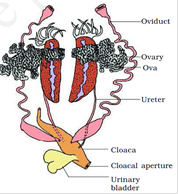
The female reproductive organs include a pair of ovaries which are situated
near kidneys. - Fertilisation is external and takes place in water. Development involves a larval stage called tadpole. Tadpole undergoes metamorphosis to form the adult.
Reproductive systems of frog-


